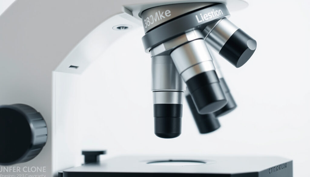
Parts of a Microscope: Functions and Components Explained
A microscope’s usefulness is decided by how accurately its mechanical frame, lenses, and illumination system work together. The parts divide into structural, optical, illumination, and adjustment roles, and microscope classes can be compared by magnification, resolution, and intended use.
Ocular lens: 10x | Objectives: 4x, 10x, 40x, 100x | Total magnification: 40x–1000x | Major components: ~15
Structural frame
- The head, arm, and base form the main frame of a compound microscope. Guyana Ministry of Education
Optical workhorse
- Objective lenses sit closest to the specimen and supply the principal magnification. Louis Pressbooks biology lab
Common powers
- School microscopes typically use a 10x eyepiece and objectives that step from 4x to 100x. Microscope.com Education Center
Magnification math
- A 10x eyepiece paired with a 10x objective produces 100x total magnification. Wiley laboratory textbook
The table below groups the most visible microscope parts by their job.
| Part | Primary function | Common source |
|---|---|---|
| Head | Supports upper optical components | Guyana Ministry of Education |
| Base | Supports the microscope and may hold the illuminator | Guyana Ministry of Education |
| Arm | Connects the head to the base and is used for carrying | Guyana Ministry of Education |
| Eyepiece | Lens viewed by the user, often 10x or 15x | Guyana Ministry of Education |
| Body tube | Keeps eyepiece and objective lenses aligned | Applied Sciences teaching notes |
| Revolving nosepiece | Holds objectives and rotates to change magnification | Guyana Ministry of Education |
| Objective lenses | Principal magnifying lenses closest to the specimen | Louis Pressbooks Biology Lab |
| Stage | Platform that supports the slide | Guyana Ministry of Education |
| Stage clips | Secure the slide when no mechanical stage is used | Microscope.com Education Center |
| Condenser | Focuses illumination onto the specimen | Louis Pressbooks Biology Lab |
What is the main function of a microscope?
The main function of a microscope is to make small structures visible by combining magnification, resolution, and illumination. A microscope that magnifies but cannot resolve detail produces a large but blurry picture.
- Magnification enlarges the image of a specimen.
- Resolution determines the ability to distinguish fine details.
- Illumination provides light to view the specimen.
How does a microscope magnify objects?
In a compound microscope, the objective lens forms a real image and the eyepiece enlarges that image a second time. The two-stage process is why the total magnification equals the objective magnification times the eyepiece magnification, as reflected in the snapshot above.
What is resolution in microscopy?
Resolution is the smallest distance at which two points can still be seen as separate. A microscope with high resolution lets you distinguish adjacent cellular structures; one with poor resolution makes them merge into a blur. Olympus Life Science explains resolution as a function of the objective lens and condenser design, not just magnification.
“The head, base, and arm are commonly identified as the microscope’s three structural parts.”
Guyana Ministry of Education
Different microscope classes handle these jobs in different ways. Light microscopes rely on visible light and glass lenses; electron microscopes use electron beams to reveal much smaller structures; confocal microscopes use laser scanning to sharpen focus in thick samples. The National Institutes of Health describes electron microscopy as a key method when resolution beyond light microscopy is needed.
The pattern: magnification enlarges, resolution clarifies, and illumination reveals; no single lens can compensate for failure in the other two.
Which lens is used in a microscope?
A standard compound microscope uses three lens systems: the objective, the eyepiece, and the condenser. The objective and eyepiece form the image; the condenser shapes the light that reaches the specimen.
What is the function of the objective lens?
Objective lenses are the primary magnifying lenses. They sit on the revolving nosepiece, closest to the specimen, and each one has a different power. The 4x, 10x, 40x, and 100x objectives are standard on many school microscopes.
What is the function of the eyepiece?
The eyepiece, or ocular lens, is the lens you look through. It magnifies the image produced by the objective and usually has a 10x or 15x power. Because the eyepiece is the last lens before your eye, it also projects the image to a comfortable viewing distance.
What is the condenser lens?
The condenser sits below the stage and focuses light into a narrow cone that passes through the specimen. A well-adjusted condenser improves both brightness and contrast. Some teaching guides describe the body tube as the part that keeps the eyepiece and objectives aligned; that alignment matters because the condenser’s light cone must meet the objective at the correct angle, as noted in Applied Sciences teaching notes.
“The eyepiece, also called the ocular, is the lens viewed by the user and commonly has 10× or 15× power in school microscopes.”
Guyana Ministry of Education
The implication: the objective does the heavy optical lifting, while the eyepiece amplifies what the objective has already resolved.
What are the three lenses of a microscope called?
The three lens systems are the objective lens, the eyepiece lens, and the condenser lens. They are named by where they sit and what they do, not by their glass alone.
What is the objective lens?
The objective lens is the lower lens group, mounted on the revolving nosepiece and positioned closest to the specimen. It provides the first stage of magnification.
What is the eyepiece lens?
The eyepiece lens is the upper lens group, mounted in the head of the microscope. It takes the image from the objective and magnifies it again for the viewer.
What is the condenser lens?
The condenser lens is the lens group below the stage. It gathers light from the illuminator and focuses it on the plane of the specimen. A microscope can still form an image without a condenser, but the image may be dim and uneven.
“Objective lenses are positioned closest to the specimen and provide the principal magnification.”
Louis Pressbooks biology lab
What this means: all three lens groups work as a system; changing one changes the character of the whole image.
What is the 100X lens on a microscope called?
The 100x lens is an oil immersion objective. To work correctly, it needs a drop of immersion oil between the lens and the slide.
What does oil immersion mean?
Oil immersion means the space between the objective lens and the cover slip is filled with oil matched to the refractive index of glass. This prevents light from bending away from the lens and lets the objective collect more of the light that passes through the specimen. Encyclopaedia Britannica describes oil immersion as a way to increase the numerical aperture and useful magnification of the objective.
What is the 10x lens called?
The 10x lens is usually called the low-power objective. It gives a wide field of view and is the best starting point for locating a specimen.
What is the 40x lens called?
The 40x lens is the high-power objective. It reveals more detail but has a narrower field of view and requires careful focus adjustment. Together, the 10x and 40x objectives cover most routine laboratory work; the 100x lens is reserved for detailed work with oil immersion.
The catch: a high numerical aperture only helps if the oil forms a continuous bridge from the lens to the slide; an air bubble can ruin the effect.
What are the two main types of eyepieces?
The two main types of eyepieces are monocular and binocular. A monocular eyepiece has one tube, while a binocular eyepiece has two tubes so both eyes can view together.
What is a monocular eyepiece?
A monocular eyepiece uses a single viewing tube. It is common on student microscopes because it is simple and inexpensive, though viewing with one eye can cause eye strain over long sessions.
What is a binocular eyepiece?
A binocular eyepiece has two viewing tubes with adjustable distance between them. It allows more comfortable viewing and helps the brain fuse the image. Many binocular heads also include a trinocular port for a camera, which makes the microscope useful for documentation.
Modern microscope heads also include built-in digital cameras and LED illumination, and manufacturers such as Zeiss Microscopy reference show how these options affect the choice of a microscope head.
Understanding how the eye and brain process visual information can help you use a microscope effectively; a Colour Blind Test NZ shows how color perception affects the same optics. Readers who want another step-by-step science explainer can follow the same structure in How to Reduce Cholesterol.
The implication: the eyepiece determines comfort and compatibility, while the objective determines how much specimen detail is actually available.
Confirmed facts and open questions
Some microscope facts are stable across teaching guides, while others depend on the source you consult.
Confirmed by multiple teaching materials
- A compound microscope uses two lens systems, the objective and the eyepiece, to form the final image. Northern Arizona University guide
- The head, arm, and base are the three structural parts named in standard school materials. Rice University teaching lab
Open questions
- The exact inventor of the compound microscope is disputed; Zacharias Janssen is often credited, but historical evidence is not conclusive.
- A lower-confidence teaching PDF says the stage opening allows illumination to pass through or around the specimen; the role is physically necessary, but the source wording is less authoritative.
The pattern: core microscope anatomy is stable across educational systems, but peripheral details such as who invented the device and how to classify every small opening can vary.
education.gov.gy, micro.magnet.fsu.edu, education.gov.gy, programs.clarendoncollege.edu
Frequently asked questions
What is a compound microscope?
A compound microscope uses two lens systems, an objective and an eyepiece, to magnify a specimen. It is the standard upright microscope used in school and clinical labs.
What is the difference between a simple and compound microscope?
A simple microscope has one lens, like a magnifying glass. A compound microscope uses two lens groups, which allows much higher magnification.
How do you calculate total magnification?
Multiply the eyepiece magnification by the objective magnification. A 10x eyepiece with a 40x objective gives 400x total magnification.
What are the focusing knobs called?
The large outer knob is the coarse focus knob; the smaller inner knob is the fine focus knob. They move the stage or body tube to bring the specimen into sharp focus.
What is the stage of a microscope?
The stage is the flat platform that supports the glass slide. Many stages include clips or a mechanical stage to hold the slide steady.
What is the purpose of the diaphragm?
The diaphragm adjusts the amount and angle of light passing through the condenser, which changes brightness and contrast.
The catch: a microscope is only as reliable as its cleanest lens and most secure slide.
For a student or lab technician, the practical result is simple: choose the objective first, adjust focus and light, and let the eyepiece confirm what the objective has resolved.
More related posts
 Christchurch to Rotorua Flights: Cheap Deals & Times
 Those Who Wish Me Dead: Plot, Cast, Reviews & Where to Watch
 Pak n Save Tauranga: Specials, Hours, Online Shopping & Jobs (Cameron Road)
 How to Connect an External Monitor to Your Laptop: Complete Guide
 The Warehouse Lower Hutt – Store Details, Hours & Online Shopping
 Windows Media Creation Tool: Download & Create Bootable USB
 Cheap Rental Cars Auckland: Budget Deals & Hidden Fees
 Inter Milan vs Arsenal FC Lineups Today
